This logo isn't an ad or affiliate link. It's an organization that shares in our mission, and empowered the authors to share their insights in Byte form.
Rumie vets Bytes for compliance with our
Standards.
The organization is responsible for the completeness and reliability of the content.
Learn more
about how Rumie works with partners.

Today, a scientist’s greatest tool for investigation may well be the microscope. If you plan on going into medicine or scientific research — or even just want to see how gnarly all the little things around you look blown up — understanding how the parts of a microscope work is a necessity. Thanks, Zacharias!
The Optical Microscope
Parts of an Optical Microscope
Let's get this bad boy out and take a look at its parts.
 Modern optical microscopes are handheld frames with a handle for carrying. The frame has two sections:
The Head
The head contains various lenses and operations for your eyes to view a sample.
The Base
The base contains a place to hold the sample. It also contains lenses and light controls.
The Microscope Head: Part I
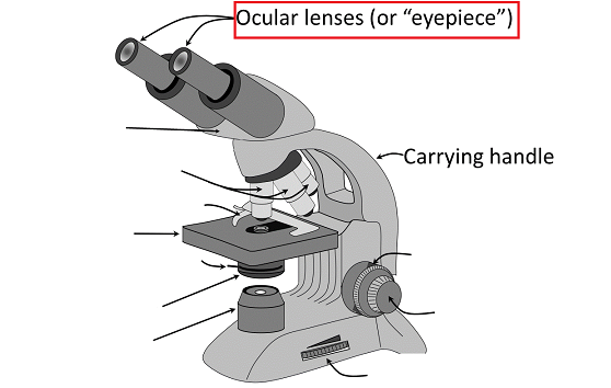
The tube that you peep through has multiple convex lenses in a stack. A convex shape just means that the glass is thick in the middle and tapered at the edges — like little glass footballs. 🏈
These tubes are called eyepieces or ocular lenses.
How it Works
Curved glass lenses increase or decrease object size when you look through them. You can see this in your daily life: in wide angle lenses in a camera 📷 — or ordinary magnifying glasses. 🔍
Where there’s light and curvy glass, size is a matter of perspective!
The Microscope Head: Part II
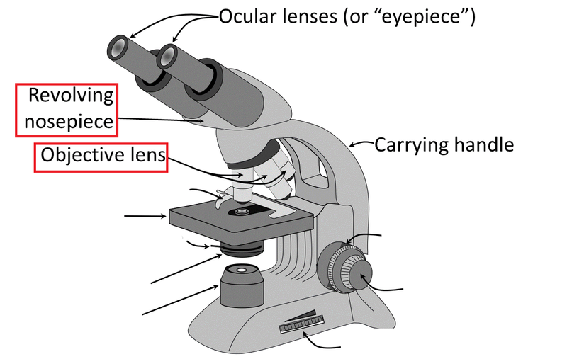
Going down the eyepiece, you’ve got more lenses closer to the object, unshockingly called objective lenses.
To increase or decrease magnification, you move the revolving nosepiece they're all attached to.
Ordinarily, you might see three objective lenses: 4x (meaning "magnified by 4 times"), 10x, and 40x.
Quiz
True or false: In general, the more lenses you stack between the eyepiece and objective lenses, the stronger your magnification will be.
Not only does piling lenses on top of each other increase magnification, but doing so usually multiplies, rather than adds, magnifying ability. You can imagine how many special ways lenses can be shaped and stacked to meet lab standards in a variety of contexts.
The Microscope Base: Part I
Don't Get Caught Off Base!
The lenses in the head are nothing without the light that passes through them. You place an interesting sample below the viewing lenses and above the light source to achieve magnification. The microscope base holds that sample and contains light controls.
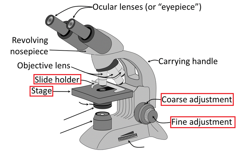
At the top of the base is the stage, which holds the sample.
The sample is placed on the stage, normally on a glass slide. It's held in place by a slide holder (also called stage clips).
Some microscopes also have a coarse and fine stage knob that raises and lowers the stage to focus the sample.
The Microscope Base: Part II
This Little Light of Mine ♪
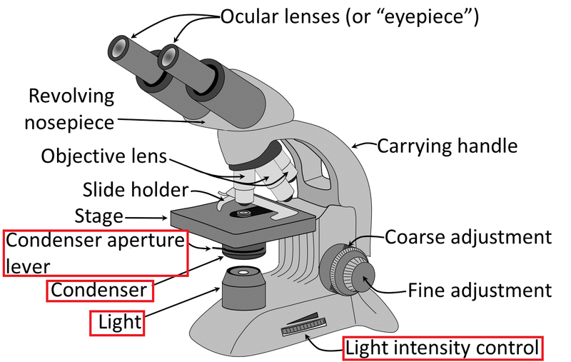
Underneath the stage is a little assembly with a big job: controlling light toward the sample.
The light intensity control gives you general control over how much light you shine toward your sample.
The light’s best friend is the condenser assembly, another collection of lenses that controls how concentrated, or condensed, the light is for your sample.
How it Works

The higher you magnify, the darker it gets! This is where the condenser literally shines: it takes the light and concentrates it into an increasingly bright cone. In general, the more you magnify, the more you’ll want to condense your light.
Quiz
You've clipped your slide in and want to see the sample at 10x zoom. But when you look through the eyepiece, everything looks dark. How might you better see the sample? Select all that apply:
Darkness is probably an issue with light, so you should work the light and condenser controls to fix it. Adjusting the stage won’t fix any light issues, and revolving the objective lenses might upset the magnification you're working in.
Did you know?
The condenser, like the eyepiece, has its own special set of lenses to help concentrate light on the sample. You may also see it called an iris or iris diaphragm.
Take Action
This Byte has been authored by
Robin Sulkosky
Composition Lecturer
MA


